Montag, 12. Oktober 2015

Prolapsed disc treatment

From the roots to the fruits: Milestones in Treatment of Prolapsed Disc

Treatment of herniated discs took his output at the beginning of the 1900s. The aim of any treatment is to find an access to the pathogenic tissue, which will reduce surrounding tissue demage in order to find a better healing process. It is a long way from the first open surgery in disc treatment to minimally invasive operation techniques that are applied today. This path is marked by milestones that have been devised by surgeons using the technological advances in medicine. The milestones are summerized in the sequence, wherein each milestone is written as an article and published as a post in "Endoscopic History"


  • Laminectomy performed by Mixter and Barr 1934
  • Postereor Cervical disc surgery performed by Spurling and Coville 1944 
  • Cervical Discectomy via anterior approach since 1950
  • Biopsie of disc material performed by Argentina 1955 and Craig 1956 
  • Chemopapain injected and discribed by Smith et al.  using the posterolateral approach 1963
  • Microdiscectomy performed and published by Caspar / Yasargil using a microscope for visualization 1975
  • Percutaneous Nucleotomy discribed and performed by Hijikata using the posterolateral approach 1975
  • Introdution of Percutaneous Nucleotomy in Europe by Schreiber, Leu, Suezawa 1978
  • Endoscopic Assisted Percutaneous Nucleotomy by Schreiber, Leu 1979
  • Development of SB-Charité lumbar disc Implant by Karin-Büttner Janz 1984
  • Percutaneous Laser Disc Decompression performed and published by Choy 1985
  • Development of legentary Minicamera MC-103 by Norbert Lemke as OEM producer for Karl Storz which influenced all endoscopic procedures since 1986 because visualization chanched from direct viewing through an Endoscope to permanent monitor contol.
  • Epiduroscopy with a 2.3 mm Micro Endoscope with working channel and angulation of the tip, published by Stoll, Watkins, Mathews 1989
  • Thoracic endoscopic acess to the spine by Mack, J.J. Regan, Rosenthal 1990
  • Biportal Approach to the spinal canal discribed and performed by Schreiber, Leu 1991
  • Foraminoscopy Presented by Hal Mathews at San Francisco Conference using a special designed Scope of  Danek Inc. 1991
  • Foraminoscopy presented by Schreiber, Leu using a special designed Scope of Karl Storz for treatment of lumbar extraforaminal herniations1991
  • Laparoscopic discectomy published by Obenchain 1991
  • Enlargment of Foramen intervertebralis with reamers and special designed instruments for removal of lumbar median, paramedian and foraminal disc herniations and treatment of stenotic foramen under C-Arm control by Thomas Hoogland 1996
  • Advanced Foraminoscopy with new designed Foraminoscope in combination with spezialized instruments instruments for lumbar median, paramedian and foraminal disc herniations and treatment of stenotic foramen by full endoscopic visualization performed by T. Hoogland 1998.
  • MED Tubular System for microdiscectomy, laminotomy, laminectomy and spinal canal stenosis published by Kevin T. Foley and Medtronic sofamor Danek 1998
  • Cervical Endoscopic Ventral Approach by Sang Ho Lee and John C.Chiu since 1998.

Mittwoch, 7. Oktober 2015

History of prolapsed disc treatment

Laminectomy

Knowledge of the lumbar herniated disc as a common cause of sciata is due William Jason MIXTER (1880 - 1958) and Joseph Seaton BARR (1901 - 1963) On September 30, 1933, the authors working at the Massachusetts General Hospital in Boston refered about  "Ruptures of the interverebral disc with involvement of the spinal canal". When etiting their 25 cases of "spinal cord tumors" they succeeded 19 times the histological detection of nucleus pulposus or anulus fibrosus and only 6 times found themselves cartilaginous or unclussifiable tumors.


Excerpt from the lecture of 1933

That, not an uncommon cause of symptoms is the herniation of the nucleus pulposus into the spinal canal or the rupture of the intervertebral disc, as we call them preferably.  The fact that the lesion was often misunderstood as cartlaginous, emananting from intervertebral disc regeneration.That in reality the rupture of the intervertebral disc ist much more commen than the neoplasia, the treatment of this disease is in our series in the 3 to 1 ratio... it is a surgical one and that the results obtained, provided that the compression was not to long, are very satisfactory".

(Mixter WJ, Barr JS (1934) Rupture of the intervertebral disc with involvement of the spinal canal. New Engl. J Med 211:210-215)

Early Operation techniques by Krause

The first actual operations of lumbar disc herniation were made in the early 1900s, the herniated discs still were called "Ekchondrome" or " Ekchondroma"also "cartilaginous tumors"
.

The type of surgical technique semms from todays´perspective adventurous (Krause 1911): Macro Surgical, so to the naked eye, the dura mater was slit-shaped opened, the cerebrospinal fluid was drained, then the intradural space was inspected, then the front of the Dura opened, and then the herniated disc was removed.

The patient, described by Krause had so far recovered from the surgery that he could lead a normal life 10 month after surgery. What remained was Peroneuspararese which could however, have also existed prior to the OP (Literature: Krause 1911, pp 717 - 719)

Operation technique by Mixter - Barr

For the laminectomy a skin incision above the center line of the spinous process is performed. The cout continous through the supraspinous ligament up to the tips of the spinous processes. There after, the spinal straps are removed, after that the lateral muscle packages are peeled off by chisel or rasp and also the lateral muscels till to the angle joints The muscels are held by a retractor aside and the spinous process is removed with a Luer forceps. Subsequently, the vertebral arches are detached with a laminectomy punch. Through removal of the spinous process and the arch the underlaying nerve was released. This operation technique has been used, with variations, till the end of the sixities last century for prolapsed disc treatment. Nowadays laminectomy is used mainly for tumors and severe spinal stenosis. Prolapsed disc is treated with more advanced operations techniques.


Sonntag, 4. Oktober 2015

Minimally invasive spinal disc treatment


Classification of proapsed spinal disc.

by intensity: Protrusion, Herniation, Sequester

by exit of the prolapsed disc: median, paramedian, foraminal, extraforaminal


by level of the prolapsed disk, cervical, thoracal, lumbal


The different prolapsed disks related to intensitiy, exit of prolaps and level require the determination of the optimized access path, operation technique as well as the appropriate instruments.

Minimally invasive disc treatment

Introduction

Herniated disc are a common musculosketeral disorder that is affected on the one hand by continuing pain and on the other hand the disease considerably restricts the living conditions. This disorder can lead to withdrawal from professional life.


The cause is the leakage of gelatinous mass from the interior of the vertebral disc. Depending on the point where the material exits, it compresses the spinal cord, the nerve root or the exiting spinal nerve, often under servere pain.As a result, it can lead to paralysis of the areas, which supplies the nerve concerned.
The exiting material has only a volume of a few cc. The problem to remedy this defect is located in the access route. The herniated disc is on one hand surrounded by sensitive structures, like spinal cord, nerves, blood vessels, ligaments and muscles, and on the other hand from a stable skeleton. This carries half of the body but also protects the neural structures.

The herniated disc occurs in very different forms, therefore the successfull treatment requires extensive preliminary investigations. These include investigations with MRI, CT and X-ray. If it is decided that after 3-6 month of unsuccessfull conservative treatment, surgery is necessary, it requires a competent surgical planning. Intensity (Protrusion, Herniation, Sequester) Level (Lumbal, thoracal, cervical) and Position (median, paramedian, foraminal, extraforaminal) have to be located and recorded. The combination of different herniated disc options by position, intensity and level require the determination of the optimized access path, the method of operation as well as the appropriate instruments.

Samstag, 30. Mai 2015

Pioneers in Endoscopy

basil hirschowitz circa1960 1


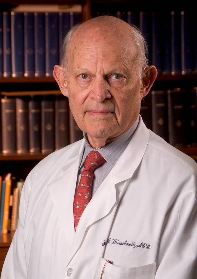
 Dr. Basil Isaac Hirschowitz

A tribute to one of the most important developers in Endoscopy, Dr. Basil Isaac Hirschowitz. We remember Dr. Hirschowitz on his birthday, 29th May. He was born 1925 and passed away 1913.

Dr. Hirschowith was a native of South Africa, born in the city Bethal. He received his medical education at the University of the Witwatersrand in Johannesburg. In 1947 Dr. Hirschowitz earned M.B and B.Ch. degrees of this institution. He received Postdoctoral training from the University of the Witwatersrand, earning an M.D degree. 1953 he moved to the United States. He continued at the University of Michigan with his gastrointestinal fellowship.

Dr. Basil Hirschowitz is best known for having invented an improved Optical Fiber Endoscope for gastroenterology. Creation of the flexible Fiber Endoscope revolutionized the practice of gastroenterology and was also a key invention in optical fiber communication in multiple industries. Never the less he used for illumination of his first flexible Fiber Endoscope a conventional light bulb.

Using fibers for illumination ( cold light) was later added by the inventors of American Cystoscope Makers and Karl Storz.

Dienstag, 7. April 2015

Endoscopic History - Founders and Inventors


Karl Storz - Founder and Inventor

In 1945, Karl Storz founded a company for manufacturing ENT instruments, headlamps, and binocular loups. This interest in medical vision, illumination, and hence physics consequently led him to spezialize in endoscopes. He was fascinated by the idea of endoscopes that allow physicians to look inside the body. At the time, the technical resources available for this purpose were still extremly limited. The examination site was illuminated using tiny, faint light bulbs, or mirrors were used to reflect external light through the endoscope tube into the body. Karl Storz quickly recognized that these methods could not achieve satisfactory illumination. He wanted to find a better way.

Two competing chracteristics benefited him in this quest: a craftsman´s sure instinct for preciese workmanship and technical feasibility on the one hand and an artist´s and inventor´s imagination on the other. His illumination solution for endoscopy is still in use today: Light generated externally by a powerful light source is transmitted into the body through a flexible fiber optic light cable. This "cold light illumination" principle laid the foundation for modern entoscopy as well as for the success of the KARL STORZ company. With more than 400 additional patents and utility models, Karl Storz has made significant impact on modern endoscopy.

(Text by Karl Storz Company delivered by Michael Künze/Karl Storz)

Montag, 6. April 2015

Endoscopic History ENT


Anton Friedrich von Tröltsch (1829 - 1890)

Anton Friedrich von Tröltsch is considered as the "Father of ENT medicine" in Germany. He was the one that added the illumination method with the reflecting mirror, developed by Hofmann (Fig. 1) into the daily clinical routine.
He gave lectures - trained by Tonybee and Wilde in England - as the first one in Germany about Otology at the Würzburg University. His published textbook of 1862 was widely circulated.
Besides his bust in the Würzburg Department of Otolaryngology and a small side street - named after him in Würzburg - reminded the angulated ear forceps (Fig. 2) to these truthfull great man of our medical field.
By Fig. 3 Tröltsch is shown during treatment of a patient. This image has been published in 1880 in a "Home Story" at the German magazine "Gartenlaube" in which he is praised in the skies. Also his presentation of the lighting mirror left downwards in the picture is of interest.


Fig. 1                                                             Fig. 2                                               Fig. 3

( Text: Dr. Lübbers, delivered by Michael Künze / Karl Storz Germany)